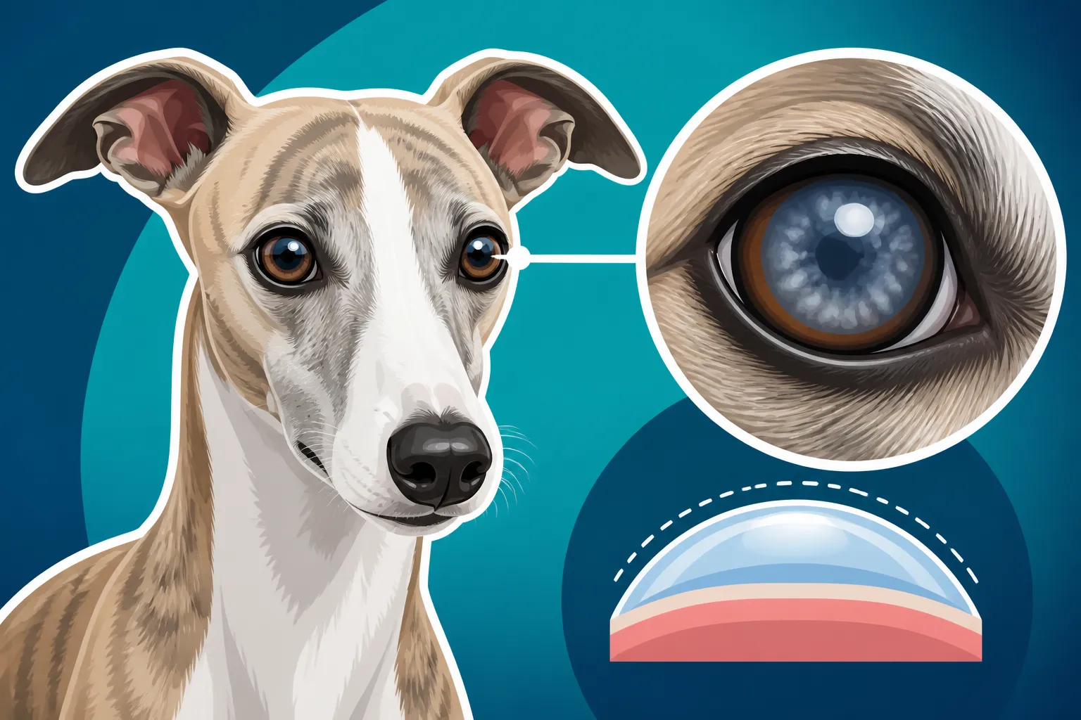
Corneal Dystrophy in a Whippet
Finding that your beloved Whippet's beautiful, soulful eyes are suddenly developing a cloudy, hazy appearance can be incredibly alarming. Your heart might sink, wondering if their vision is being affected, or if something much more serious is going on. This guide is here to walk you through understanding corneal dystrophy in Whippets, helping you identify the signs, manage the condition, and ensure your lean, elegant companion continues to see the world as clearly and comfortably as possible.
Understanding Corneal Dystrophy in Whippets
Corneal dystrophy is one of those veterinary terms that sounds intimidating, but at its core, it refers to a non-inflammatory, inherited condition that affects the cornea of the eye. Unlike an injury or infection, which would cause an inflammatory response, corneal dystrophy is a metabolic defect that causes abnormal deposits in one or more layers of the cornea. It's not usually painful in its early stages, but it can certainly be confusing and worrisome for us pet parents.
The Cornea and Its Role
To truly understand corneal dystrophy, it helps to know a little about the cornea itself. Think of the cornea as the clear, protective "front window" of your Whippet's eye. It’s a transparent, dome-shaped tissue that covers the iris, pupil, and anterior chamber, playing a crucial role in focusing light onto the retina. Its clarity is paramount for good vision. The cornea has three main layers:
- The Epithelium: This is the outermost protective layer, constantly regenerating.
- The Stroma: The thickest middle layer, responsible for the cornea's strength and transparency. It's mostly collagen fibers arranged in a very organized way.
- The Endothelium: The innermost layer, a single layer of cells that pumps fluid out of the cornea to keep it clear and prevent swelling.
Corneal dystrophy can affect any of these layers, leading to different types of the condition and slightly varied appearances in your Whippet's eye.
Different Types and What They Look Like
While corneal dystrophy is a general term, it typically manifests in a few different forms, primarily categorized by which layer of the cornea is affected. For Whippets, the most commonly discussed form is stromal dystrophy, although epithelial and endothelial types can also occur.
- Epithelial Dystrophy: This type affects the outermost layer. You might notice small, usually bilateral, white or gray dots or lines, often centrally located. These can sometimes lead to recurrent erosions (small, painful ulcers) because the epithelial cells don't adhere properly. It’s less common in Whippets than stromal dystrophy.
- Stromal Dystrophy: This is the big one for Whippet owners. It's characterized by grayish-white, crystalline, or sparkling opacities (cloudiness) within the middle layer of the cornea. These deposits are typically made of lipids (fats) or cholesterol and can appear as rings, dots, or diffuse haziness. They usually affect both eyes and are often symmetrical. The good news is that while they can look quite dramatic, these stromal opacities rarely cause significant vision impairment because they are usually non-progressive or progress very slowly, and are often located in areas that don't block central vision entirely.
- Endothelial Dystrophy: This type affects the innermost layer responsible for keeping the cornea clear of fluid. When these cells fail, the cornea can swell (edema), leading to a bluish-white cloudiness. This is often more serious as it can cause significant vision impairment and discomfort. Endothelial dystrophy is generally seen in older dogs and specific breeds like Boston Terriers or Chihuahuas, and is less common in Whippets compared to stromal dystrophy.
Here's a quick overview of the main types and what you might observe:
| Type of Dystrophy | Affected Layer | Typical Appearance | Common in Whippets | Impact on Vision |
|---|---|---|---|---|
| Epithelial | Outermost | Small white/gray dots, lines; can cause erosions | Less Common | Minimal, but erosions can be painful and impair |
| Stromal | Middle | Grayish-white, crystalline opacities; symmetrical | Most Common | Usually minimal to moderate, often slowly progressive |
| Endothelial | Innermost | Bluish-white corneal swelling (edema) | Less Common | Can be significant, especially as it progresses |
It's vital to remember that these descriptions are generalizations. Only a veterinary ophthalmologist can truly diagnose the specific type of corneal dystrophy affecting your Whippet.
Spotting the Signs: What to Look For
The earliest signs of corneal dystrophy can be incredibly subtle, which is why keeping a close eye on your Whippet's general health, including their eyes, is so important. Many owners first notice something when their Whippet is between two and five years old, though it can appear earlier or later. You might be petting them, or looking at them from a particular angle, and suddenly spot something that wasn't there before.
Visual Changes in the Eye
The most obvious symptom of corneal dystrophy is a change in the appearance of one or both eyes. This isn't usually a sudden, dramatic event, but rather a gradual development.
- Cloudiness or Haziness: This is often the first thing people notice. It can look like a faint, milky haze, or more distinct white or grayish spots within the clear part of the eye. For stromal dystrophy, these might even have a crystalline or "sparkling" quality, almost like tiny sugar crystals under the surface.
- Bilateral Involvement: While it might appear in one eye first, corneal dystrophy is almost always bilateral, meaning it affects both eyes, though not always to the same degree or at the same time. If you only see changes in one eye, it's more likely to be an injury or infection, which needs immediate veterinary attention.
- Symmetrical Appearance: In many cases of stromal dystrophy, the pattern of opacities can be surprisingly similar in both eyes. This symmetry is a strong clue for dystrophy rather than other eye conditions.
- Location of Deposits: The deposits often appear in the central or paracentral (just off-center) region of the cornea.
When you notice these changes, try to take a clear picture in good lighting to show your vet. It can be hard to describe accurately, and a visual aid helps them assess the progression.
Behavioral Cues and Discomfort
One of the reassuring aspects of many corneal dystrophies, particularly stromal dystrophy, is that they often don't cause significant pain or discomfort in their early stages. However, as the condition progresses or if complications arise, your Whippet might start showing signs that indicate irritation.
- No Pain, Initially: Unlike corneal ulcers or foreign bodies, uncomplicated corneal dystrophy isn't usually painful. Your Whippet typically won't be squinting, holding the eye shut, or tearing excessively at first. This can be misleading, making owners delay a vet visit, but it's important to get it checked out regardless.
- Increased Blinking or Squinting: If the dystrophy causes the corneal surface to become irregular, or if it leads to secondary issues like epithelial erosions, your Whippet might start blinking more frequently or squinting. This indicates irritation.
- Rubbing at the Eye: You might notice your Whippet pawing at their face, rubbing their eye against furniture, or on the carpet. This is a clear sign of discomfort and needs to be investigated, as rubbing can exacerbate any underlying issue or even cause further damage.
- Excessive Tearing or Discharge: While not typical for uncomplicated dystrophy, if the condition makes the eye more prone to irritation or leads to secondary infection, you might see increased watery discharge or even thick, colored discharge.
- Vision Changes: While stromal dystrophy often has minimal impact on vision, especially early on, extensive opacities can certainly reduce your Whippet's ability to see clearly. You might notice them bumping into things in low light, hesitating on stairs, or being less sure-footed, particularly if the opacity is central. Given that Whippets are sighthounds, their vision is paramount, and even subtle changes can affect their confidence. For other eye conditions that can lead to blindness, you might want to read about Whether a Whippet Goes Blind From Progressive Retinal Atrophy.
It's easy to dismiss a subtle eye change, especially if your Whippet isn't acting uncomfortable. But with any eye issue, it's always best to err on the side of caution and get a professional opinion.
Diagnosis and Management of Corneal Dystrophy
When you notice any changes in your Whippet's eyes, the first step is always a visit to your regular veterinarian. They can perform an initial assessment and, if they suspect corneal dystrophy or another complex eye issue, will likely recommend a consultation with a veterinary ophthalmologist. These specialists have advanced equipment and expertise to properly diagnose and manage eye conditions.
The Diagnostic Process
Diagnosing corneal dystrophy usually involves a thorough ophthalmic examination. This is quite different from a regular check-up and can be fascinating to watch.
- Thorough Ophthalmic Exam: The vet will use an ophthalmoscope and often a slit lamp biomicroscope to examine all parts of the eye, including the eyelids, conjunctiva, cornea, lens, and retina. A slit lamp allows them to examine the individual layers of the cornea in detail, which is crucial for identifying the exact location and nature of the deposits.
- Fluorescein Stain: A fluorescein dye is applied to the eye to check for corneal ulcers or abrasions. This dye sticks to any areas where the outermost epithelial layer is damaged, glowing bright green under a special light. In uncomplicated corneal dystrophy, the stain will typically not stick, indicating that the surface of the cornea is intact.
- Ruling Out Other Conditions: It's critical to differentiate corneal dystrophy from other conditions that can cause similar-looking cloudiness, such as corneal ulcers, keratitis (corneal inflammation, often due to injury or infection), corneal edema from glaucoma or uveitis, or even fatty deposits (lipid keratopathy) that are secondary to systemic issues like hypothyroidism or high cholesterol. This is why a full diagnostic workup is so important. Sometimes, blood tests might be recommended to rule out underlying systemic diseases.
Once a diagnosis of corneal dystrophy is made, your vet or the ophthalmologist will discuss the specific type and the best management plan for your individual Whippet.
Treatment and Management Options
It's important to understand that there is no "cure" for corneal dystrophy as it is an inherited condition. The goal of management is to minimize symptoms, prevent complications, and maintain your Whippet's comfort and vision.
- No Treatment Often Needed for Mild Cases: For many Whippets with mild stromal dystrophy, especially if it's not progressing rapidly and isn't impacting vision or causing discomfort, active treatment might not be necessary. Regular monitoring by an ophthalmologist is usually sufficient. This might involve yearly check-ups, which are also a good idea for general eye health, as discussed in Whether a Whippet Needs a Yearly Eye Exam.
- Lubricating Eye Drops: If the eye feels dry or slightly irritated due to the dystrophy, over-the-counter lubricating eye drops or artificial tears can be used to keep the corneal surface moist and comfortable. Your vet can recommend suitable options.
- Hyperosmotic Agents: For some forms, especially those involving fluid accumulation or more pronounced stromal deposits, hyperosmotic saline drops or ointments (e.g., Muro 128) might be prescribed. These work by drawing excess fluid out of the cornea, helping to improve clarity. These are available by prescription and should only be used under veterinary guidance.
- Addressing Secondary Issues: If the dystrophy leads to complications like recurrent corneal erosions or ulcers, these will need to be treated aggressively with topical antibiotics to prevent infection, and sometimes pain medication. It's not uncommon for a Whippet's The Thin Skin That Sends a Whippet to the Vet for Stitches to be delicate, and eye issues can be similarly sensitive.
- Surgical Intervention (Rare): In very severe, vision-impairing cases, surgical options like superficial keratectomy (removing the superficial layers of the cornea) might be considered. However, this is typically a last resort and often the deposits can reoccur. Surgery also carries risks and a significant recovery period, and it's essential to weigh the potential benefits against these factors. Most ophthalmologists prefer medical management for corneal dystrophy.
When dealing with specialist visits and potential long-term medication, it's wise to consider the financial implications. Pet insurance, like a plan from Healthy Paws, can be a real lifesaver when unexpected health issues like corneal dystrophy arise, helping to cover the costs of diagnostics, specialist consultations, and ongoing treatments.
Living with Corneal Dystrophy: Long-term Care and Prognosis
Discovering your Whippet has corneal dystrophy can be a bit disheartening, but it’s crucial to remember that for most dogs, especially Whippets with stromal dystrophy, the prognosis for a good quality of life and preserved vision is generally excellent with proper management. This is not a death sentence, but rather a condition that requires your attentive care and collaboration with your veterinary team.
Daily Care and Monitoring at Home
Your role as a pet parent is paramount in managing corneal dystrophy and preventing complications. Integrating eye care into your daily routine is often quite simple.
- Consistent Application of Eye Drops: If your vet prescribes drops or ointments, consistency is key. Set reminders and ensure you apply them as directed. Positive reinforcement and treats can make this a more pleasant experience for your Whippet. Gently wiping away any excess with a clean tissue can also prevent build-up around the eye.
- Regular Eye Inspections: Make it a habit to look at your Whippet's eyes daily. Look for any changes in cloudiness, signs of redness, discharge, or if they seem to be squinting or rubbing their eyes more than usual. Early detection of secondary issues can prevent them from becoming more serious.
- Environmental Management: While not a direct treatment, ensuring your Whippet's environment is comfortable can help. Minimize exposure to dust, strong winds, or irritants that could dry out their eyes or cause secondary issues. If they love running through brush, for instance, be extra vigilant about checking their eyes afterward. This kind of careful daily attention is part of What a Whippet Needs Every Day for overall health.
- Keeping Up with Vet Appointments: Adhering to the schedule set by your general vet or ophthalmologist for follow-up appointments is critical. These visits allow them to monitor the progression of the dystrophy, adjust treatments if necessary, and catch any secondary issues early.
Potential Complications and When to Worry
While many cases of corneal dystrophy are benign, it's important to be aware of potential complications that do require immediate veterinary attention.
- Recurrent Corneal Erosions/Ulcers: This is one of the most common complications, particularly with epithelial dystrophies, but can also occur if the corneal surface is compromised by other types. An ulcer is a painful open sore on the cornea. Signs include intense squinting, tearing, redness, and pawing at the eye. If you see these signs, contact your vet immediately.
- Vision Impairment: While often minimal, significant or rapidly progressing cloudiness can reduce your Whippet's vision. If you notice them struggling to navigate familiar environments, especially in low light, or appearing startled by objects, it's time for a vet check.
- Pain and Discomfort: While initial dystrophy is not painful, any sign of pain, such as holding the eye shut, light sensitivity, or rubbing, indicates a problem that needs to be addressed. It could be an ulcer, an infection, or another painful complication.
Don't hesitate to contact your vet if you notice any sudden worsening of symptoms or signs of discomfort. It's always better to be safe than sorry when it comes to eye health. The cost of veterinary care can accumulate, so reviewing your pet insurance policy, understanding What a Whippet's Annual Deductible Should Be, and ensuring coverage for specialist visits can provide significant peace of mind.
Prognosis and Quality of Life
For most Whippets with corneal dystrophy, especially the common stromal type, the prognosis for long-term comfort and good vision is excellent. The condition is rarely progressive to the point of causing blindness and often requires minimal intervention beyond monitoring. Many Whippets live out their full The Twelve to Fifteen Years a Whippet Lives with corneal dystrophy without significant impact on their active, happy lives.
However, it's a condition that emphasizes the importance of responsible breeding practices. Since corneal dystrophy is often inherited, breeders should be mindful of the health of their breeding stock. While for a pet owner the focus is on management, for the broader breed, considering Whether a Whippet Should Be Screened Before Breeding can help reduce the prevalence of such conditions.
Ultimately, living with a Whippet who has corneal dystrophy means being a diligent observer and a proactive advocate for their eye health. With your love and consistent care, your Whippet will continue to be the bright-eyed, cherished member of your family they've always been.
Disclaimer
The information provided in this article is for educational purposes only and is not a substitute for professional veterinary advice. Always consult with a qualified veterinarian or veterinary ophthalmologist for any health concerns or before making any decisions related to your Whippet's care.